Mi Band®
February 29, 2024
AI Technology Semi-Supervised Machine Learning
March 1, 2024AI Improves Pediatric, Ophthalmologic, and Dermatologic Monitoring

AI helps pediatricians check heart health.
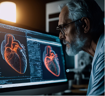
Pediatric cardiologist Charitha Reddy, MD, recognized the need for a faster and more reliable method to assess the heart’s pumping ability in children, a crucial indicator of cardiac health. Collaborating with engineers and computer scientists, Reddy developed an AI model tailored to pediatric patients, automating the estimation of the left ventricle’s function. The model demonstrated superior speed and consistency by analyzing thousands of heart ultrasound videos and generating accurate assessments of heart-pumping capacity compared to human evaluations. Published in the Journal of the American Society of Echocardiography, the study highlights the model’s potential to enhance efficiency in medical decisions, particularly for children undergoing treatments like chemotherapy. Reddy envisions broader applications, including remote screenings for heart conditions in underserved areas, pending further testing and validation of the model’s performance.(1)

Furthermore, Reddy’s AI model opens doors for future advancements in pediatric cardiology, potentially extending its capabilities to assess the function of the right ventricle and evaluate fetal hearts or those with structural abnormalities. With continued refinement and testing, this innovative technology holds promise in improving diagnostic accuracy and efficiency and expanding access to specialized cardiac care for children, ultimately enhancing health outcomes and quality of life. (1)
Pediatric cardiologist Charitha Reddy, MD, recognized the need for a faster and more reliable method to assess the heart’s pumping ability in children, a crucial indicator of cardiac health. Collaborating with engineers and computer scientists, Reddy developed an AI model tailored to pediatric patients, automating the estimation of the left ventricle’s function. The model demonstrated superior speed and consistency by analyzing thousands of heart ultrasound videos and generating accurate assessments of heart-pumping capacity compared to human evaluations.

Published in the Journal of the American Society of Echocardiography, the study highlights the model’s potential to enhance efficiency in medical decisions, particularly for children undergoing treatments like chemotherapy. Reddy envisions broader applications, including remote screenings for heart conditions in underserved areas, pending further testing and validation of the model’s performance.Furthermore, Reddy’s AI model opens doors for future advancements in pediatric cardiology, potentially extending its capabilities to assess the function of the right ventricle and evaluate fetal hearts or those with structural abnormalities. With continued refinement and testing, this innovative technology holds promise in improving diagnostic accuracy and efficiency and expanding access to specialized cardiac care for children, ultimately enhancing health outcomes and quality of life. (1)
AI analyzed repurposed chest CT images to identify calcium buildup in arteries, encouraging patients to make lifestyle changes.

Cardiologist Alexander Sandhu has encountered numerous patients who could potentially benefit from the diagnostic insights provided by a computed tomography (CT) scan of their cardiac structures to discern the presence of calcium buildup within their coronary arteries. Such plaque accumulation serves as a robust predictor of susceptibility to heart attacks. However, a considerable portion of patients forego this diagnostic procedure, often due to limitations in insurance coverage. Nevertheless, Sandhu identified a fortuitous circumstance several years ago wherein many patients had previously undergone chest CT scans for unrelated reasons. Recognizing the potential inherent in repurposing these scans to extract pertinent cardiac information, Sandhu contemplated employing artificial intelligence (AI) algorithms to glean valuable insights from such scans, initially conducted for disparate medical purposes, such as lung cancer screening.(2)
This unexpected observation prompted Sandhu, in collaboration with his mentor, Dr. David Maron, Director of Preventive Cardiology at the School of Medicine, to embark on the development of a deep learning algorithm tailored to extract meaningful cardiac data from chest CT scans. They aimed to achieve accuracy comparable to dedicated cardiac CT scans specifically ordered to assess coronary calcium. Conducting initial trials in 173 high-risk patients for heart disease, the team observed promising results. Upon notifying patients and their primary care physicians about the presence of coronary calcium detected through the algorithm’s analysis, there was a notable increase in the initiation of statin medications within six months, as detailed in Sandhu’s subsequent analysis published in the journal Circulation in November 2022. This proactive approach to identifying asymptomatic patients at risk of cardiovascular events underscores the potential of AI-driven methodologies to enhance preventive cardiology practices.(2)

Cardiologist Alexander Sandhu has encountered numerous patients who could potentially benefit from the diagnostic insights provided by a computed tomography (CT) scan of their cardiac structures to discern the presence of calcium buildup within their coronary arteries. Such plaque accumulation serves as a robust predictor of susceptibility to heart attacks. However, a considerable portion of patients forego this diagnostic procedure, often due to limitations in insurance coverage. (2)
Nevertheless, Sandhu identified a fortuitous circumstance several years ago wherein many patients had previously undergone chest CT scans for unrelated reasons. Recognizing the potential inherent in repurposing these scans to extract pertinent cardiac information, Sandhu contemplated employing artificial intelligence (AI) algorithms to glean valuable insights from such scans, initially conducted for disparate medical purposes, such as lung cancer screening.This unexpected observation prompted Sandhu, in collaboration with his mentor, Dr. David Maron, Director of Preventive Cardiology at the School of Medicine, to embark on the development of a deep learning algorithm tailored to extract meaningful cardiac data from chest CT scans. They aimed to achieve accuracy comparable to dedicated cardiac CT scans specifically ordered to assess coronary calcium. Conducting initial trials in 173 high-risk patients for heart disease, the team observed promising results. Upon notifying patients and their primary care physicians about the presence of coronary calcium detected through the algorithm’s analysis, there was a notable increase in the initiation of statin medications within six months, as detailed in Sandhu’s subsequent analysis published in the journal Circulation in November 2022. This proactive approach to identifying asymptomatic patients at risk of cardiovascular events underscores the potential of AI-driven methodologies to enhance preventive cardiology practices.(2)
Google is rolling out new AI models for healthcare.
Google has launched MedLM, a suite of health-care-specific artificial intelligence models to assist clinicians and researchers in complex studies and doctor-patient interaction summaries. This move signals Google’s latest endeavor to monetize AI tools in the healthcare sector amid fierce competition from industry rivals like Amazon and Microsoft. The MedLM suite comprises large and medium-sized AI models built on Med-PaLM 2, a language model trained on medical data. While the suite is now available to eligible Google Cloud customers in the U.S., Google also plans to introduce health-care-specific versions of its Gemini AI model to MedLM in the future. (3)

The real-world application of MedLM includes aiding emergency medicine physicians in automatic documentation of patient interactions and streamlining nurse handoffs, showcasing potential efficiency gains. However, challenges such as incorrect information outputs and user intent deciphering limitations highlight the need for cautious implementation and ongoing refinement.(3)
Google has launched MedLM, a suite of health-care-specific artificial intelligence models to assist clinicians and researchers in complex studies and doctor-patient interaction summaries. This move signals Google’s latest endeavor to monetize AI tools in the healthcare sector amid fierce competition from industry rivals like Amazon and Microsoft. (3)

The MedLM suite comprises large and medium-sized AI models built on Med-PaLM 2, a language model trained on medical data. While the suite is now available to eligible Google Cloud customers in the U.S., Google also plans to introduce health-care-specific versions of its Gemini AI model to MedLM in the future.The real-world application of MedLM includes aiding emergency medicine physicians in automatic documentation of patient interactions and streamlining nurse handoffs, showcasing potential efficiency gains. However, challenges such as incorrect information outputs and user intent deciphering limitations highlight the need for cautious implementation and ongoing refinement.(3)
AI better photos of skin for telehealth visits in medicine
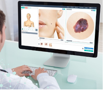
When the COVID-19 pandemic necessitated a shift to telehealth at Stanford Medicine’s dermatology clinic in 2020, Roxana Daneshjou, MD, Ph.D., grappled with the challenge of interpreting patient-submitted photos of skin conditions. Recognizing the potential for artificial intelligence (AI) to streamline this process, Daneshjou and her team embarked on developing an algorithm to assess and enhance the quality of these images automatically. Stanford’s Catalyst program secured funding to create TrueImage, a web app designed to guide patients in capturing clinically useful photos using their smartphones or tablets. The app, which identifies and rejects low-quality images while guiding improvement, has shown promising results in pilot studies, reducing the submission of poor-quality photos by 68%. With further clinical trials underway to validate its efficacy, TrueImage holds the potential to expedite dermatology appointments nationally and assist healthcare providers beyond dermatology in collecting quality images for diagnostic purposes. (4)

When the COVID-19 pandemic necessitated a shift to telehealth at Stanford Medicine’s dermatology clinic in 2020, Roxana Daneshjou, MD, Ph.D., grappled with the challenge of interpreting patient-submitted photos of skin conditions. Recognizing the potential for artificial intelligence (AI) to streamline this process, Daneshjou and her team embarked on developing an algorithm to assess and enhance the quality of these images automatically. Stanford’s Catalyst program secured funding to create TrueImage, a web app designed to guide patients in capturing clinically useful photos using their smartphones or tablets. (4)

The app, which identifies and rejects low-quality images while guiding improvement, has shown promising results in pilot studies, reducing the submission of poor-quality photos by 68%. With further clinical trials underway to validate its efficacy, TrueImage holds the potential to expedite dermatology appointments nationally and assist healthcare providers beyond dermatology in collecting quality images for diagnostic purposes.o.(1,2,3)
An AI model could predict whether patients need eye surgery to prevent vision loss.

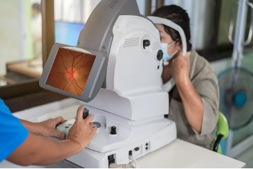
Glaucoma specialist Dr. Sophia Wang, alongside her collaborators, recognized the challenge of identifying which patients with glaucoma would progress rapidly and require surgical intervention to prevent irreversible blindness. To address this, they explored the potential of artificial intelligence (AI) to analyze doctors’ notes and clinical measurements to predict the need for surgery among glaucoma patients. Training a two-pronged AI model on data from thousands of glaucoma patients, they found that the model could accurately assess a patient’s likelihood of needing surgery within a year with over 80% accuracy. This surpasses the predictive abilities of even highly trained specialists like Dr. Wang herself. While further refinement and testing are necessary, incorporating imaging data and validating the model across diverse patient populations will be crucial to its potential clinical application in guiding treatment decisions for glaucoma patients. (5)

Glaucoma specialist Dr. Sophia Wang, alongside her collaborators, recognized the challenge of identifying which patients with glaucoma would progress rapidly and require surgical intervention to prevent irreversible blindness. To address this, they explored the potential of artificial intelligence (AI) to analyze doctors’ notes and clinical measurements to predict the need for surgery among glaucoma patients. (5)
Training a two-pronged AI model on data from thousands of glaucoma patients, they found that the model could accurately assess a patient’s likelihood of needing surgery within a year with over 80% accuracy. This surpasses the predictive abilities of even highly trained specialists like Dr. Wang herself. While further refinement and testing are necessary, incorporating imaging data and validating the model across diverse patient populations will be crucial to its potential clinical application in guiding treatment decisions for glaucoma patients.(5)
